Scanning electron microscopy (SEM)
With the scanning electron microscope (SEM) we visualize surface structures of organisms. Beside a better resolution, it also provides a larger depth of field than a binocular microscope.
A fine electron beam, controlled by deflection coils, illuminates the sample pixel by pixel. The interaction of the primary electron beam with the sample surface leads to the emission of secondary electrons, backscattered electrons and X-ray radiation that are collected by the related detectors.
For generating surface images, primarily the secondary electrons are used. Backscatter electrons and X-ray emission are used for investigating the elemental composition of a sample (see section > Spectroscopic Methods<).
As the whole system, the sample is placed in high vacuum (10-5 - 10-7 mbar). Therefore it has to be completely dried to not affect the vacuum. Dehydration takes place with chemical or physical techniques. A special case is the use of fresh, undried samples in environmental mode that indeed is limited in resolution in view of the required low vacuum in the sample chamber (about 1 mbar).
We moreover offer a novel method to transfer hydrated samples immediately in the SEM and observe them in high resolution, the cryo transfer. This is also useful for samples that produce severe shrinking artifacts by drying them (eg. mosses, water plants or hydrogels). Samples will be rapidly frozen in liquid nitrogen and can be directly introduced into the high vacuum on a cooled cryo stage.
A further benefit is the possibility to break open samples to expose internal structures. Remaining cell water has to be sublimated by heating up to -90°C, which we call freeze etching.
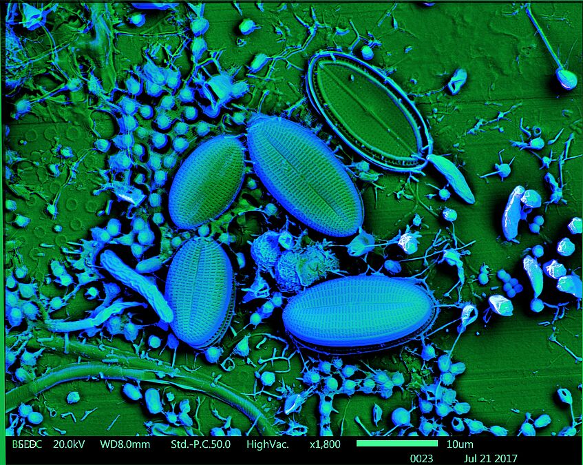
limnetic biofilm, artificially colored (Daniela Gruber)

limnetic biofilm, artificially colored (Daniela Gruber)