Light Microscopy
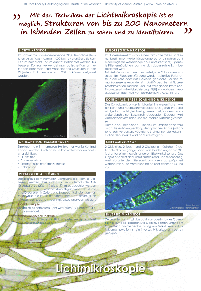
Microscopy is our main tool to investigate cells and their contents. Routinely, we use all contrast methods in light microscopy like differential interference contrast, phase contrast, dark field and polarisation. Submicroscopical structures are visualised in fluorescence and confocal laser scan microscopy. Due to our focus on dynamic processes in living cells, we are also well equipped with video and film techniques for documentation and analysis through the microscope.
> Light microscopical techniques
> Fluorescence microscopy
> Video and UV microscopy
> Confocal laser scanning microscopy
Electron Microscopy
In order to visualize submicroscopic structures, we provide scanning-(SEM) and transmission electron microscopes (TEM) as well as associated equipment for sample preparation. In addition, we are equipped for spectrometric techniques (EDX, EELS) to study the elemental composition of biological samples. We also adapt and refine cryo fixation and dehydration methods for various tissue types.
Electron microscopes surpass the resolution of visible light which is limited to about 300 nm (wavelength/2). Electrons can be excited to much higher energies, corresponding to shorter wave lengths. This enables spatial resolutions of about one nanometer. The electrons are emitted by a cathode, accelerated by high voltage (1-300 kV), deflected by electromagnetic lenses (coils) and limited by apertures to create a controllable beam.
This advantage of SEM and TEM (high resolution, possibility for element analyses) has to be paid with higher technical effort (need of dry samples, high vacuum, and extensive sample preparation).
> Scanning electron microscopy (SEM)
> Transmission electron microscopy (TEM)
Scientific Documentation
Scientific Documentation
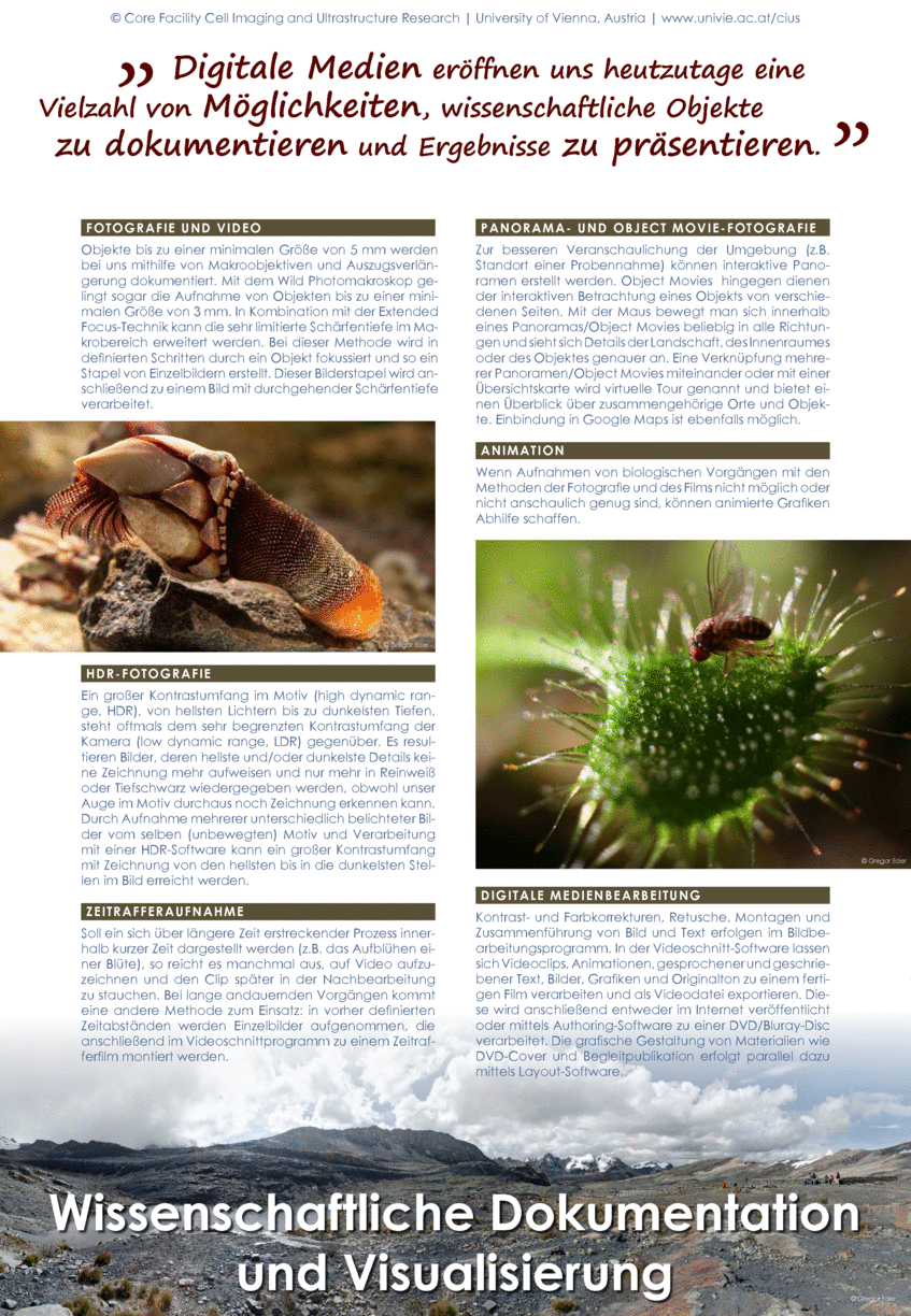
With our scientific documentation and visualization tools we do macro photography and video, HDR photography, time lapse photography, panorama and object movie photography, animations and media design.
Micromanipulation and microinjection
Micromanipulation and microinjection
Micromanipulation and microinjection can be used to induce local pressure and wounding of cells. Furthermore, specific dyes and drugs can be injected into single cells to analyze the consequences within the injected cell and its neighbours. Intracellular ion measurement also uses the micromanipulation equipment and shows a cell`s reaction to external stress according to changes in the ion content.
The above methods require very skillful handling and a lot of practice. In our laboratory, micromanipulation and microinjection techniques can be conveniently combined with video equipment to monitor dynamic processes and reorganizations of cells and their contents.