Transmission electron microscopy (TEM)
Transmission electron microscope (TEM) is used to visualize thin sections of resin embedded samples. Samples has to be fixed to >freeze< or >immobilize< the current metabolic status. This takes place by chemical treatment or physically by quick-freezing the samples under high pressure (see Cryo-methods).
Fixed samples are dehydrated by organic solvents and embedded in synthetic resin (Epoxid, Araldit, LR White, …). Ultrathin sections (about 50 nm thin) will be made with a diamond knife on an ultramicrotome and mounted on a TEM-grid, consisting of copper, zinc or gold.
In the TEM column a constant laminar electron beam transmit the sample and display an electron density profile, which is converted to photons in the scintillator and read out as a digital image of grey values. The lower the electron density of a material, the brighter is the area of the image.
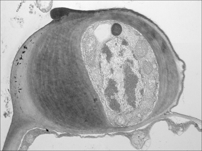
Avena sativa, primary stomata cell (Marieluise Weidinger)
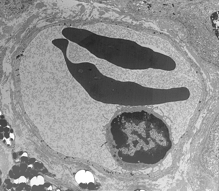
Plethodon shermani, blood vessel showing a lymphocyte and two erythrocytes (Norbert Cyran)
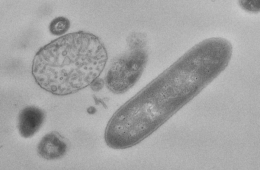
Bacteria from coffee machine deposit (Norbert Cyran)